2026
Sup252
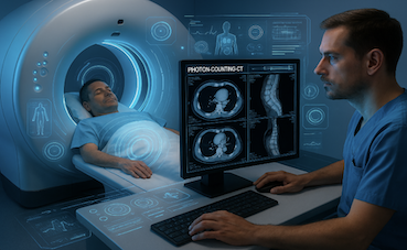
CT Image Storage for Processing
This Supplement adds For Processing storage SOP Classes based on the
existing CT Image IOD, Enhanced CT Image IOD, and Legacy Converted
Enhanced CT Image IOD.
For Processing storage SOP Classes in DICOM facilitate the exchange
and storage of images needed for processing while distinguishing them
from those for presentation.
This supports appropriate data pipelines while not disrupting reading
workflow with images not intended for presentation.
These new SOP Classes mirror existing Mammography, Intra-Oral X-ray,
and Digital X-ray For Processing SOP Classes.
One application of the For Processing SOP Classes is to store and
exchange CT basis images created by the multi-energy decomposition
process.
These are not typically diagnostic themselves, but can be processed to
generate an extensive variety of diagnostic images (iodine maps,
virtual non-contrast images, virtual monoenergetic images at various
energy levels, calcium maps, etc.).
Hanging Protocols would typically ignore these For Processing
images.
This supplement was voted ready to be incorporated into the standard
as Final Text in publication 2026c.
View slideset »
2025

Sup249
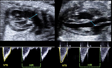
Ultrasound Fetal Anatomy Survey SR Extensions
This supplement to the DICOM Standard introduces new SR
template content to address fetal anatomy survey
assessments in ultrasound reports.
Specifically, a sub-template is added to TID 5000 along
with corresponding CIDs to address the anatomy of interest
and assessments for each.
Clinical guidelines from the International Society of
Ultrasound in Obstetrics and Gynecology (ISUOG) call for a
survey of fetal anatomy in the first, second, and third
trimesters to identify structural anomalies.
In Japan, JSUM guidelines call for first and second
trimester anatomical surveys
The guidelines identify specific lists of anatomy to
consider.
This supplement was voted ready to be incorporated into
the standard as Final Text in Publication 2025e.
View slideset »

Sup247
Eyecare measurements templates
This Supplement proposes to add templates, context groups,
and coded vocabulary for eyecare measurements to the
Standard.
These templates may be used in either SR documents, or for
structured content in an Encapsulated PDF object.
The focus of this Supplement is the set of “key”
measurements clinically important for patient care. These
are not intended to be a comprehensive set of ophthalmic
measurements, although the extensible context groups and
templates allow additional measurements beyond the
specified key measurements to be included in SOP
Instances.
The key measurements of this Supplement are primarily
derived from analysis of images, in particular retinal
optical coherence tomography (OCT) images. Note that there
are several existing IODs that record measurements
directly produced by various refractive devices that do
not produce images (autorefraction, lensometry,
keratometry, etc.), as well as more comprehensive visual
field and macular thickness reports, which are not
intended to be replaced by these more summary key
measurement templates.
There is tension in clinical documentation between the
needs for structured discrete data and human-readable
content. In DICOM, discrete data is generally sent using
Structured Reporting, and ready for display rendered data
may be sent in an Encapsulated PDF.
A given set of measurements may be sent in objects in both
formats, with cross-reference to the other object using
the Referenced Instance Sequence (0008,114A); note that
the cross-reference is to an instance as a whole, not to
individual measurements. Alternatively, discrete
measurements may be included in an Encapsulated PDF object
in the SR-like Content Sequence (0040,A730).
The Templates defined in this Supplement may be used in
either object type.
The DICOM Standard does not recommend the use of any
particular approach to meeting the clinical documentation
needs of the users.
Such recommendation may be made by a professional society
or a standards profiling effort. For example, the American
Academy of Ophthalmology and the IHE Eyecare domain,
considering the need to integrate legacy PDF-based
systems, have in the past recommended use of Encapsulated
PDF with the included SR-like Content Sequence for basic
interoperability, but those recommendations may not meet
all use cases in the evolving interoperable healthcare IT
environment.
This supplement was voted ready as Final Text and to be
incorporated into the next publication of the standard
(2025c).
View slideset »
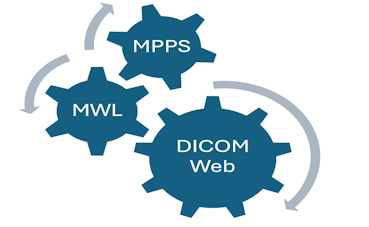
Sup246
DICOMweb Modality Procedure Step Services
This supplement adds the Modality Scheduled Procedure Step
Service and the Modality Performed Procedure Step Service
to DICOMweb to mirror the Modality Worklist (MWL) and
Modality Performed Procedure Step (MPPS) services that are
already available in DIMSE respectively.
The modality procedure step services have been designed
with the intention of facilitating proxies from/to
DIMSE.
This supplement was voted ready as Final Text and to be
incorporated into the next publication of the standard
(2025d).
View slideset »

Sup244
Frame Deflate Transfer Syntax
This Supplement adds a new Transfer Syntax primarily for
single bit segmentation encoding, which is otherwise not
well supported.
There is a need to be able to store and transfer encoded
single frames (such as for DICOMweb) rather than the
entire dataset for those applications where only selected
frames of a multi-frame object are required (such as for
selected tiles at selected resolutions for whole slide
images that have been segmented, or multi-organ
segmentations of large volumetric CT or MR
datasets).
Currently, the DICOM standard supports a means of single
bit representation of binary segmentations with a Bits
Stored and Bits Al- located of 1, and these can grow
extremely large, especially when segmenting at the full
resolution of the underlying image (e.g., for whole slide
imaging).
If compressed, they need to be mathematically reversibly
(losslessly) compressed. The existing Deflate Transfer
Syntax (algorithm used in zip and gzip) is reasonably
effective, but applies to the entire data set (including
the "metadata" and all the frames treated as a single
stream).
Frame-based pixel data compression schemes currently in
the standard generally do not support single-bit, with the
exception of RLE and J2K (CP 2301), neither of which
achieves as high a compression ratio as Deflate does for
segmentation data.
Other alternative lossless compression codecs designed for
single bit use (such as for fax using CCITT Group 4 (ITU-T
T.6), JBIG, or JBIG2) were considered, which though they
compress more effectively, were not considered widely
enough supported to justify the complexity for this use
case at this time. Other general purpose compressors do
slightly better than Deflate, but again, not so much
better that they justify their addition to the standard at
this time, though they may be considered in future if
other use cases justify them.
This supplement was voted ready for Final Text and
publication in the 2025a edition of the standard.
View slideset »

Sup241
Structural Heart SR Template
This supplement introduces SR templates for
Structural Heart Procedures.
These procedures involve interventions aimed at
addressing various conditions or abnormalities
affecting the structures of the heart, excluding the
coronary arteries.
Unlike open-heart surgery, these interventions are
characterized by their minimally invasive nature or
catheter-based approach.
Periprocedural imaging follows a consistent pattern
of three phases: pre-operative assessment,
intraprocedural assessment, and follow-up.
Throughout all three phases, echocardiography
emerges as the primary imaging modality.
X-ray angiography is predominantly utilized for
intraprocedural guidance.
CT may also find application in the pre-operative
assessment and follow-up.
The templates proposed in the supplement are based
the Simplified Adult Echocardiography Templates
(root TID 5300), modified to support multimodality
image acquisition.
Structural Heart Procedures include:
- TAVI: Transcatheter Aortic Valve Implantation
- TAVR: Transcatheter Aortic Valve Replacement
- TTVr: Transcatheter Tricuspid Valve Replacement
- TTVR: Transcatheter Tricuspid Valve Repair
- TEER: Transcatheter Edge-to-Edge Repair
- TMVR/TMVr: Transcatheter Mitral Valve Replacement
- LAAO: Left Atrial Appendage Occlusion
This supplement was voted ready for Final Text and to be incorporated into the next publication of the standard (2025b).
View details »
View slideset »
Sup236
Waveform Presentation State
This supplement introduces Service Classes for
storage and exchange of presentation information for
DICOM waveform objects by adding a Waveform
Presentation State IOD. The Waveform Presentation
State object stores the display montages,
i.e. calculative combinations of recorded channels,
display filter and other display properties as well
as arbitrary Annotations.
This supplement adds
- a new Waveform Presentation State IE.
- a SOP Class to store Waveform Presentation States and the related Modules.
In cardiology, technicians annotate previously recorded waveforms (e.g. from home monitoring Holter ECG) and highlights areas of interest. This information is essential input for the cardiologist who reviews the ECG and finally provides the report.
This supplement was voted ready for Final Text and publication in the 2025a edition of the standard.
View details »
View slideset »

Sup233
Patient Model Gender Enhancements
This supplement adds a comprehensive gender logical model for sex and
gender representation in DICOM.
This facilitates communication between DICOM and the
various HL7 systems.
The goal is to make the distinction between phenotypical
sex and the patient's social context gender clear.
Adds optional attributes to the Patient Study Module and
to various C-FIND and normalized services. These optional
attributes match those in the HL7 logical model.
The DICOM model extensions are consistent with the work in
HL7 and FHIR:
- The HL7 Gender Harmony Project created a logical model to describe
the information needed in an electronic record to support proper
care for gender and sex diverse patients.
- The HL7 model includes both clinical information and
social information.
This supplement also updates Patient Sex (0010,0040) description and
some CIDs to match the HL-7 updated definition.
This supplement was voted ready for Final Text and to
be incorporated into the next publication of the
standard (2025b).
View slideset »
2024
Sup243
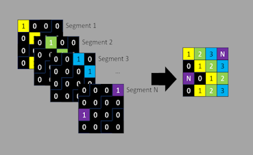
Label Map Segmentation
This Supplement describes addition of a Label Map
Segmentation IOD to DICOM to encode classification of
entities.
Currently, the DICOM standard supports an IOD and SOP
Class for pixel- or voxel-based segmentation encoding (as distinct
from the representation of segmented objects as surfaces in the
surface segmentation and encapsulated 3D object IODs and SOP Classes),
in which each segmented property is represented as a binary bit plane
(or an 8 bit probabilistic or occupancy value).
While this allows for overlapping of segments, it is
inefficient and difficult to encode large numbers of
non-overlapping segmentations, as they require non-trivial
processing both to extract from the bit plane encoded
data, to assure there is no overlap, and to convert to the
label map form that is very commonly used internally and
persistently for clinical applications.
The current DICOM bit-plane-based segmentation methods
have proven to be awkward both for 3D cross-sectional
imaging applications when there are very large numbers of
slices and/or structures, and for whole slide microscopy
imaging, when there are very large numbers of tiles and/or
property classes.
They are also typically large and sparse and should
compress well but there are very few single bit
compression schemes supported by the standard and they do
not do well with these types of images.
This Supplement defines a label map segmentation enhanced
multi-frame IOD that specifies a data structure that
provides, for each pixel or voxel in 2D, 3D or tiled
pyramidal space, an index value conveying the
non-overlapping segment for each pixel.
Existing data elements for describing segmentations are
reused where appropriate.
Bit depth is sufficient (8, 16) to encode large numbers of
segments but allow for more compact encoding.
The existing palette color photometric interpretation may
be used (instead of monochrome) if colors are to be
suggested, to leverage the widespread implementations in
toolkits, and to allow for the use of existing lossless
com- pression schemes.
Segment properties are conveyed in the existing segment
description structure so as to be compatible with the
existing bit plane segment descriptions.
Re-using the segment description does not prevent the use
of separately encoded or well- known DICOM color palette
objects.
The scope is confined to label maps for "classes" (what
"class" a segment represents) but not "instances' (which
"instance" of a "class" is represented), where classes and
instances are separately communicated by the pixel value
(e.g., if one wants to individually identify nuclei rather
than treat them all as being of one class).
This might be the subject of a future extension.
The scope is confined to a single label map, which does
not allow for overlap of different segments.
If overlapping of multiple label maps is required,
separate SOP Instances may be created.
Issues related to the efficient representation (or
avoidance) of the Per-Frame Functional Group Sequence (in
which, for every frame, the Referenced Segment Number is
specified) are out of scope, and may be addressed in a
separate Supplement or CP if necessary.
This supplement was voted ready as final text and is
incorporated in publication 2024d.
View slideset »
Sup242
Ultrasound Fetal Cardiac SR Extensions
This supplement to the DICOM Standard introduces new SR
template content to address fetal cardiac assessments in
echo reports.
Current clinical practice and technology for fetal cardiac assessments
using ultrasound have progressed since Sup78 was published, which
introduced TID 5220 "Pediatric, Fetal and Congenital Cardiac
Ultrasound Reports" and sub-template TID 5228 "Cardiac Ultrasound
Fetal Measurement Section".
Practice now includes many more measurements beyond visual
assessment. For example, additions will address:
- measurements of the ventricles, atria, septa and valves,
- measurements of fetal arrhythmia and hemodynamics,
- assessment of the fetal cardiovascular profile score (CVPS)
Many measurements described for pediatric echo are also potentially relevant for fetal echo, particularly at later stages of fetal development.
To that end, TID 5221 is now included in TID 5228, making any of those measurements readily available as needed and appropriate.
Also, CID 12279, which is titled Cardiac Ultrasound Fetal General Measurement, is pruned here based on usage experience to list just general fetal measurements that are specifically relevant to cardiac fetal ultrasound.
CID 12005 Fetal Biometry Measurement already covers fetal measurements relevant to a non-cardiac fetal ultrasound. Since CID 12279 is extensible, any existing implementations with unexpected usages will not be invalidated.
This supplement was voted ready as final text and is incorporated in publication 2024d.
View details »
View slideset »

Sup240
Heightmap Segmentation
This Supplement introduces a new Heightmap Segmentation IOD and SOP
Class.
Heightmaps in computer graphics are defined as a two-dimensional
raster image used to store surface elevations that can later be
applied to a three-dimensional object.
In its DICOM use, heightmap is a type of segmentation using a 2D set
of pixels to identify a surface in the 3D volume of a referenced
multi-frame image.
In the degenerate case, it can identify the intersection of a surface
with a single image plane, i.e., a 1D raster for a 2D object.
The Heightmap Segmentation IOD follows the current enhanced
multi-frame image data architecture.
For data management purposes, e.g., with Media Exchange, Heightmap
Segmentation SOP Instances may be treated similarly to other
segmentation images.
While intended to be broadly applicable for a variety of medical
imaging domains, the initial use case is in ophthalmic tomography
(OPT) for representing segmentation of retinal layers.
Further description of Heightmap Segmentation is found in the proposed
informative annex to PS3.17.
This Supplement also revises the current Ophthalmic Optical Coherence
Tomography En Face Image IOD, which had required use of Surface
Segmentation SOP Instances to specify a retinal layer, to allow use of
any type of segmentation SOP Instances, including Heightmap
Segmentation or other (including future) SOP Classes.
The reference to the segmentation object in the En Face Image object
enables traceability of the processing steps that produced the
image. It is not necessarily the case that a receiving application
could reproduce the En Face Image from the original source Ophthalmic
Tomography Image(s) and the referenced segmentation object(s).
This supplement was voted ready as final text and is
incorporated in publication 2024d.
View slideset »
Sup239
Waveform Annotation Structured Report
This supplement introduces SOP Classes for storage and
exchange of waveform annotations. It applies to all
modalities in which waveform objects are created and
applications used to review them.
Waveform Annotations annotations can be stored in the
waveform object itself expressing physical or
environmental circumstances noted by the recording device
at recording time.
The new IOD can be used to store additional clinical
information added at recording time or later provided
either by a human reviewer (for example a neurologist or a
technologist) or by an automated analysis software.
This supplement:
- Adds a SOP Class to store observations and measurements in a Waveform Annotation SR.
- Defines a new Root Template derived from TID 1500, a waveform analogy to TID 1600 Image Library, and some included templates to store annotations as codes or free text and 90 measurements.
- Defines the Context Groups used in these Templates.
View details »
View slideset »

Sup234
DicomWeb Storage Commitment
This supplement adds storage commitment
functionality to DICOMweb.
This is an extension to the existing DICOMweb
services, mimicking the storage commitment service
that is already available using DIMSE.
The storage commitment service is typically used
when an image acquisition system wants to free up
storage space for new studies and asks an archive
system of taking over the storage responsibility for
the images previously being sent from the
acquisition device to the archive.
A design goal for this supplement is to relatively
easy create proxies for Storage Commitment with
combinations of DIMSE and DICOMWeb
communication.
The DICOMweb variant of Storage Commit extends the
DIMSE variant.
In DICOMweb it is possible to provide the study and
series context to the referenced instances; this
provides more information for finding these
instances at the server side.
This supplement is incorporated into the standard as
Final Text in the publication 2024a.
View slideset »
Sup232
JPEG XL
This supplement adds lossless, JPEG recompression and
general JPEG XL Transfer Syntaxes.
JPEG XL has the following desirable features:
- JPEG XL has demonstrated improved compression of color images
- Existing Baseline JPEG images can be transcoded without additional loss to smaller JPEG XL images (particularly useful for WSI)
- Supports multi-frame encoding more effectively than animated gif, the only other multiframe rendered format
- JPEG XL has both lossless and lossy modes that can be natively displayed in some browsers
- Has flexible encoding options (including > 8 bits, single bit)
JPEG XL is also added to the set of rendered formats for DICOMweb.
- It avoids the need to transcode into JPEG
- Performance is adequate even with WASM based decoders
This supplement was voted ready as final text and is incorporated in publication 2024d.
View details »
View slideset »
Sup228
DICOMweb API for Server Volumetric Rendering
This supplement introduces Volumetric Rendering web
services and a Volumetric Rendering Protocol IOD to
enable Volume Rendering (VR), Maximum Intensity
Projection (MIP), and Multiplanar Planar Rendering
(MPR) without having to specify the numerous and
complex parameters required to do so.
Web services enable a user agent to initiate
server-side 3D volumetric rendering by specifying
Query Parameters and/or referencing a Volumetric
Rendering Protocol, or a Volumetric Presentation
State.
The Resources introduced in the Supplement derive
Query Parameters from Volumetric Presentation State
attributes while maintaining alignment with current
DICOMweb Studies Rendered Resources.
The Volumetric Rendering Protocol IOD is a
Non-Patient Instance within the Defined Procedure
Protocol IOD family.
Its primary function is to facilitate the creation
of predefined renderings, by establishing criteria
and organizing image set inputs for rendering, and
specifying Volumetric Rendering parameters, such as
rendering algorithms, geometry, color, shading, and
lighting.
This supplement was voted ready as final text and is
incorporated in publication 2024d.
View slideset »
2023
Sup237
General 32-bit ECG Waveform
This supplement defines a new ECG Waveform SOP Class
(based on the existing General ECG SOP Class) with
fewer constraints.
This High-Resolution SOP class permits 32 bits per
sample. The already available General ECG SOP
class can store waveform with 16 bits per sample.
In clinical neurophysiology it is common practice to
acquire ECG data together with the routine scalp EEG
or in case of a sleep study.
This supplement was voted ready to go out for Letter
Ballot review and voting.
This supplement was voted ready as Final Text and will
be incorporated into the standard in the next
publication (2023c).
View slideset »
Sup235
High-Throughput JPEG 2000 (HTJ2K) Compression
This supplement adds the HTJ2K Transfer Syntax to
Part 5.
HTJ2K speeds-up JPEG 2000 by an order of magnitude
at the expense of slightly reduced coding
efficiency.
HTJ2K retains JPEG 2000's advanced features, with
reduced quality scalability, while being faster and
much more efficient than traditional JPEG.
This is achieved by replacing the Part 1 block coder
with an innovative block coder for today's
vectorized computing architectures.
Detailed information is available at
jpeg.org.
This supplement is voted ready as Final Text and
will be incorporated in publication 2023e.
View slideset »
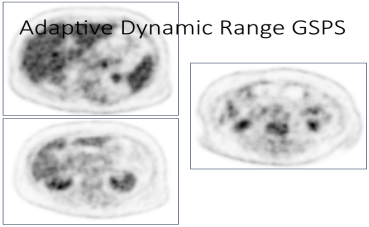
Sup231
Variable Modality LUT Softcopy Presentation State
This supplement defines a new Variable Modality LUT
Softcopy Presentation State SOP Class for both
grayscale and pseudo-color.
This new SOP class differs from existing SOP classes
in that it allows the Modality LUT to be controlled
for each image or frame.
This is intended for modalities in which the dynamic
range varies between images or frames, resulting in
each referenced image having a different Modality
LUT.
In DICOM, Presentation States are intended to be a
complete specification of the presentation to
provide consistent presentation.
An aspect of this is that PS3.4 N.2.1.1 requires the
Modality LUT in the image be ignored in the presence
of a GSPS object, even if no Modality LUT is
explicitly defined in the GSPS. Further, the GSPS
only supports one Modality LUT.
This is problematic in cases such as PET or MR, in
which the dynamic range of the measured values
varies between images.
Without this new SOP Class, the GSPS creator would
be forced to render multiple GSPS objects, one for
Modality LUT change.
This supplement was voted ready as Final Text and
will be incorporated into the standard in the next
publication (2023b).
View slideset »
Sup229
Photoacoustic Imaging
This Supplement introduces a new IOD and a new
storage SOP Class for encoding and storing
photoacoustic images.
Photoacoustic (PA) imaging enables imaging optical absorption in
biological tissues with acoustic resolution.
Contrast is generated through absorption by chromophores that range
from intrinsic absorbers such as hemoglobin and melanin to extrinsic
agents such as indocyanine green (ICG) or diverse types of
nanoparticles.
In principle, excitation at multiple wavelengths allows the modality
to discriminate individual chromophores.
Prospective applications in the space of clinical imaging range from
classification of breast cancer lesions through screening of sentinel
lymph nodes to assessment of inflammation.
Photoacoustic Imaging is in widespread use in preclinical research
labs and is currently being translated to clinical applications in
first commercial implementations.
Many (but not all) PA implementations integrate active pulse/echo
ultrasound in a hybrid imaging system to capitalize on
well-established contrast for anatomical information.
The scope of this IOD is to define the a Photoacoustic (PA) images and
processed images that may be derived from a combination of these PA
images. Complementary images such as pulse/echo ultrasound are
represented by their native DICOM IODs.
Albeit fusing PA images with US images for display is the presently
most common scenario, the particulars of the fusion are beyond the
scope of this IOD, but examples are provided.
PA images represent image output generated by the input of one or more
optical excitation wavelengths. PA images may result from excitation
by light pulses at one or more wavelengths.
A closely related but out of scope imaging modality
is Thermoacoustic imaging (TAI) which uses microwave
radiation to excite the tissue (in contrast to light
pulses).
This supplement was voted ready as Final Text and
will be incorporated into the standard in the next
publication (2023c).
View slideset »
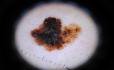
Sup226
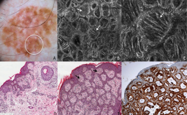
Confocal Microscopy
This Supplement to the DICOM Standard introduces two
new IODs (Confocal Microscopy IOD, Confocal
Microscopy Tiled Pyramidal Image IOD).
These IODs are intended to be applicable to all
application of confocal microscopy.
An acquisition context module specific to cutaneous
confocal microscopy is defined.
Cutaneous confocal microscopy is a non-invasive
imaging technique that allows examination of the
skin at resolutions comparable to histology without
performing biopsy.
Cutaneous confocal microscopy may be done in-vivo or
on ex-vivo tissue.
In-vivo cutaneous reflectance confocal microscopy
(RCM) is used for the early diagnosis of a range of
cutaneous diseases with an emphasis on melanoma and
pigmented lesions.
In-vivo cutaneous RCM is most often used as an
adjunct to clinical and dermoscopic imaging of a
skin lesion as opposed to a stand-alone imaging
technique.
In addition to diagnostic applications, in-vivo
cutaneous RCM may be used for the pre-operative
mapping of margins of ill-defined tumors, which
allows more accurate surgical plan and reduces
surgical morbidity.
The cutaneous RCM microscope uses a diode laser as a
source of monochromatic and coherent light and
scanning and focusing optical lens to penetrate the
skin and illuminate a small tissue spot. Reflected
light forms an image on a photodetector.
Ex-vivo cutaneous confocal microscopy allows the
microscopic examination of freshly excised
tissue.
The ex-vivo cutaneous confocal microscopy can work
in reflectance mode or fluorescence mode.
When using the fluorescence mode, the entire
surgical specimen is dipped in a solution of a
fluorescent agent and subsequently rinsed to remove
excess of fluorescent agent.
This supplement is voted ready as Final Text and
will be incorporated in publication 2023e.
View slideset »
2022
Sup230
TLS Security Update 2021
This Supplement adds two new Secure Transport
Connection Profiles and retires several others.
The IETF recently updated the Best Current Practice
document called BCP-195. The new document no longer
allows downgrading to TLS 1.0 or 1.1, which
necessitates DICOM retiring Secure Transport
Connection Profiles that are based on those
protocols.
The new version of BCP-195 is more in
line with DICOM's B.10 Non-Downgrading BCP 195
Secure Transport Connection Profile.
In addition, the Japanese government has modified
their guidelines for "high-security type" devices,
hence the old Extended BCP 195 profile (B.11) is
also now out of date, needs to be retired, and a new
profile created that reflects the new revisions.
Supplement 230 was voted to be incorporated into the standard as Final Text in publication 2022e.
View slideset »
Sup227
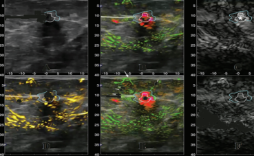
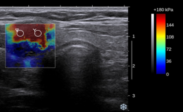
Elastography SR Template
This supplement to the DICOM Standard introduces
an SR section template for Ultrasound Elastography results
and a General Ultrasound Report within which it can be
used.
Ultrasound elastography is used on tissues including
liver, breast, prostate, and tendon. In shear wave
elastography (SWE), the ultrasound system measures shear
wave speed (SWS) and derives a value for elasticity (in
kPa) from that.
Some systems also assess viscosity (which can be
correlated to inflammation) by generating a value such as
shear wave dispersion slope.
In strain elastography (SE), elasticity/stiffness is
assessed qualitatively by comparing the compression of
tissue in a target region to that of tissue in a nearby
reference region.
This supplement is added into the standard in publication
2022c.
View slideset »

Sup225
Multi-Fragment Video Transfer
This supplement adds new video transfer syntaxes to remove
the restriction that the video cannot be broken up into
multiple fragments.
The affected transfer syntaxes are:
- MPEG2 Main Profile / Main Level Video Compression
- MPEG2 Main Profile / High Level Video Compression
- MPEG-4 AVC/H.264 High Profile / Level 4.1 Video Compression
- MPEG-4 AVC/H.264 High Profile / Level 4.2 Video Compression
A significant motivation for this supplement is the appearance of videos of size larger than 2^32-2 bytes (eg procedure video recordings).
Supplement 225 was voted to be incorporated into the standard as Final Text in publication 2022b.
View details »
View slideset »
Sup223
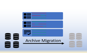
Archive Inventory
This Supplement introduces a new Repository Query SOP
Class to obtain an inventory of a repository system, a
composite Inventory IOD that is the equivalent persistent
instantiation of such an inventory, an Inventory Creation
SOP Class to initiate asynchronous creation of Inventory
SOP Instances, and SOP Classes to transfer, query and
retrieve Inventory SOP Instances.
There are considerable use cases for these new services:
Porting large DICOM repositories from one image management
system (PACS or VNA) to another.
Migration approaches need to operate at large scales, and
handle both on-premises and remote (e.g., cloud-based)
storage.
Migration often occurs when either the source system or
the destination, or both, are in clinical operation, but
systems designed and configured to handle the throughput
of regular operations might not have capacity for the
additional massive input/output requirements of migration.
Healthcare institutions merge previously disparate
repositories into an enterprise repository.
Research use cases, including artificial intelligence and
machine learning, where bulk access to the archive is
desirable, and such uses might leverage some of the same
mechanisms developed for migration.
PACS audit and quality control may also utilize some of
the standardized functionality developed for migration,
such as an archive inventory and metadata to identify the
data produced by a particular unit or by a particular
modality.
A key requirement for migration (and other use cases) is
the ability to have an inventory of all studies, series,
and instances from an archive.
This Supplement specifies a new Repository Query SOP Class
that includes features supporting a sequential set of
queries intended to produce a complete repository
inventory. These features include well-defined behavior
for queries that reach a system limit for number of
responses, and an ability to resume at the next record in
a subsequent query.
The current Query Service (DIMSE or equivalent DICOMweb)
has limitations on number of responses and the synchronous
protocol require the use of a possibly very large number
of partial query requests, with undefined behavior when
query limits are exceeded.
This Supplement also specifies an Information Object
Definition capable of encoding an inventory of all
studies, series, and instances in a repository. This is
functionally equivalent to a query response that returns
an inventory of the entire repository database, or a
subset thereof as specified by key attributes.
The Supplement further defines a mechanism to remotely
initiate the production of the inventory through a DICOM
network service and allow production to proceed
asynchronously.
Only inventory of patient-related studies, series and
instances is defined. Inventory of non-patient objects is
out of scope for this Supplement.
This supplement is not in itself a complete standard for
migration.
This supplement is added into the standard in publication
2022c.
View slideset »
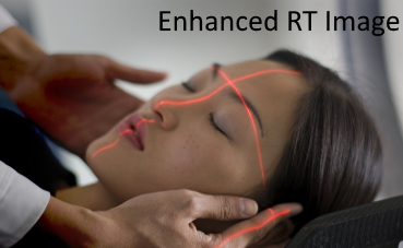
Sup213
2G-RT: Enhanced RT Image
The Supplement addresses imaging within Radiotherapy treatment
sessions and acquiring patient positioning information.
The supplement adds three IODs. Two for supporting projection images
and one IOD supporting acquisition instructions for images and other
artifacts to be used for patient positioning.
The Enhanced RT Image covers the images with a smaller number of
frames, where the per-frame functional group macros are populated for
all frames.
The Enhanced Continuous RT Image covers images which are continuously
acquired, resulting in high number of frames due to a high frame
rate. With frame level attributes not being repeated for each frame
this image type is more efficiently and sparsely populated.
Both IODs represent projection images of the patient geometry in
relation to the treatment device equipment. They may be used to guide
the positioning of the patient in respect to the treatment delivery
device to ensure delivery of the therapeutic dose to the intended
region. They may also be used to verify the position of the patient
when acquired prior, during or after the delivery of the therapeutic
radiation.
The Supplement additionally specifies a new IOD to convey parameters
instructing devices on how to acquire images or other artifacts used
for patient position verification in Radiotherapy treatment delivery
sessions.
RT Patient Position Acquisition Instruction contains the definition of
the procedures, devices, and related parameters to be used for the
assessment and/or verification of the patient position. The technical
parameters can be defined on any level of detail as needed by a
specific device.
Procedures can be paired to represent related operations like e.g. a
paired orthogonal MV and kV image acquisition.
The scope of therapeutic radiation whose position is verified is
specified by referencing SOP Instances identifying objects like RT
Radiation Set IOD of RT Radiation IODs.
Supplement 213 was voted to be incorporated into the standard as Final Text in publication 2022e.
View slideset »

Sup209
Revision of DICOM Conformance Statement
This Supplement provides updates to part PS3.2 of the
DICOM standard, redefining the content and structure of
the DICOM Conformance Statement to better meet the needs
of all user groups, for example service, R&D, testing,
sales, healthcare provider IT personnel.
Comparability is better facilitated for different products' DICOM
functionality by providing essential information in tables.
Ambiguities and inconsistencies will be less frequent between
different vendor documentations.
Web services and security are additionally addressed in the
conformance statement.
A detailed template is provided. Vendors are encouraged to populate
this template for their products. Template-based comparison of
products is advantageous in many situations.
Supplement 209 was voted to be incorporated into the standard as Final Text in publication 2022e.
View slideset »
2021

Sup222
Whole Slide Imaging Annotation
This Supplement to the DICOM Standard specifies a new
DICOM Information Object and Storage SOP Class for storing
Microscopy Bulk Simple Annotations (points, open
polylines, closed polygons and simple geometric shapes
without relationships), which is referred to as the
Microscopy Bulk Simple Annotations IOD.
Microscopy Bulk Simple Annotations are usually created by
machine algorithms from high resolution images of entire
tissue sections, e.g., encoded as DICOM Whole Slide
Microscopy images.
These annotations are distinct from
alternative representations appropriate for different
use-cases, such as segmented bit planes (which are encoded
in DICOM Segmentation Images), and more tractable size
human or machine generated contour-based annotations on
selected high-power fields or lower resolution or gross
specimen images (which are encoded in DICOM Structured
Reports using standard templates like TID 1500).
No new image encoding mechanism is introduced. The
annotations are either 2D image relative (frame or Total
Pixel Matrix) or in a 3D Frame of Reference that is shared
with a Microscopy Image Storage instance.
No new composition mechanism is added. The annotations are
basic ("simple") and it is anticipated that in future
mechanisms such as the Radiotherapy Conceptual Volume
mechanism may be re-used to describe boolean
relationships, etc., that reference instances of bulk
simple annotations, or embed more complex relationships.
Supplement 222 was voted to be incorporated into the standard as Final Text.
View slideset »
Sup220
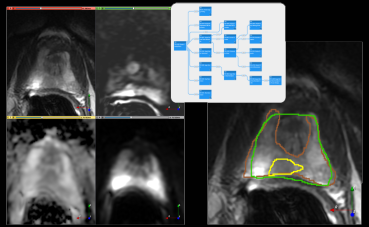
MR Prostate Imaging Structured Report
This supplement to the DICOM standard introduces a DICOM
Structured Reporting template for encoding the
radiologist's interpretation of a patient's prostate MR
imaging study.
The primary purpose of the templates is to support linking
of the annotations of findings with the measurements
derived from those findings and qualitative evaluations
associated with those findings.
In addition, the template provides the means to
communicate image-related information that is important to the
assessment of prostate MRI (e.g., Prostate Specific Antigen testing
history and prostate biopsies).
The main use cases motivating the development of the
templates are the following:
- Interoperability between the radiology workstations used for
annotation of prostate MRI and MRI-Ultrasound fusion and targeting
workstations used for sampling suspected locations using targeted
biopsy approaches;
- Interoperability with the machine learning tools that automate the
process of identifying suspected prostate cancer locations;
- Collection of structured data to support training of the machine
learning tools for automated detection and grading of prostate cancer
in MRI;
- Aggregation of structured MRI interpretation documents across
institutions;
- Integration of structured machine-readable information annotating clinical findings in MRI longitudinally and across radiology, urology and pathology subspecialties.
While the organization of the templates is not restricted to a specific prostate MRI interpretation protocol, it is primarily designed to support Prostate Imaging - Reporting and Data System (PI-RADS), which is a prostate MRI structured reporting and scoring guidelines developed through an international collaboration of the American College of Radiology (ACR), European Society of Uroradiology (ESUR), and AdMetech Foundation.
Supplement 220 was voted to be incorporated into the standard as Final Text. View details »
View slideset »
Sup214
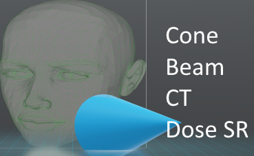
Cone-beam CT Radiation Dose SR
This supplement creates a new DICOM SR IOD with the
necessary flexibility to address cone-beam CT (CBCT)
acquisitions.
CBCT is used in multiple fields (e.g., dentistry,
radiotherapy, interventional radiology, image guided
surgery), and there are different methodologies for
describing the dose associated with each application
(typically borrowing from either XA or CT).
However, the underlying data acquisition, reconstruction,
and testing parameters for image quality and dose
evaluation are similar.
The proposed supplement defines a generic framework for
the description of radiation dose amongst the different
CBCT applications.
It retains the capability to store legacy dosimetric
values (e.g., CTDI, DAP), while allowing for reduced
dependence on modality-specific conditions for populating
fields.
This generic radiation description is capable of
representing acquisition types that already exist in the
standard (Angiography, Mammography, CR/DR, CT).
There are two fundamental inclusions in the proposed supplement:
- Decoupling of irradiation events and dose descriptions
(allowing dose-related characteristics to span multiple
irradiation events, or breaking irradiation events into
smaller time periods). For characteristics that remain
constant (e.g., focal spot size), a value can be encoded
once for the 90 entire SR. For characteristics that change
within irradiation events (e.g., tube current), multiple
values can be encoded for improved understanding of dose
distributions.
- Improved geometric description of the system. Describe the spatial relationship of different system components with respect to one another for modeling of spatial distributions of dose.
Radiotherapy treatment dose and radiopharmaceutical dose are out of scope.
Supplement 214 was voted to be incorporated into the standard as Final Text. View details »
View slideset »
Sup212
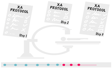
XA Protocol
This Supplement defines a pair of storage SOP Classes to distribute
defined XA protocols and to record performed XA protocols.
The two storage SOP Classes are:
- XA Defined Procedure Protocol Storage SOP Class that describes desired values (and/or value ranges) for various parameters, which includes acquisition, reconstruction and storage tasks. Defined Protocols are independent of a specific patient. Defined Protocols are typically specific to a certain acquisition equipment model and/or version (identified by device attributes in the protocol), but model-non-specific protocols are not prohibited.
- XA Performed Procedure Protocol Storage SOP Class that describes the values actually used in a performed procedure. Performed protocols are patient-specific.
The SOP Classes address details including:
- patient preparation & positioning
- equipment characteristics
- acquisition technique
- reconstruction technique
- preliminary image handling such as filtering, enhancement
- results data storage (auto-sending)
The primary goal is to set up the acquisition (and reconstruction) equipment, not to script the entire behavior of the department, or the angiographic suite. The protocol object supports simple textual instructions relevant to the protocol such as premedication, patient instructions, etc. Formal coding and management of instructions may be handled with other objects and services such as the Contrast Injection SR or the Modality Worklist (MWL).
It is expected that the vast majority of protocol objects will be specific to a certain model and version of acquisition equipment. There is no requirement that an equipment be able to run a protocol from another equipment.
Supplement 212 was voted ready as Final Text. It is incorporated into the standard in the update "2021a". View details »
View slideset »
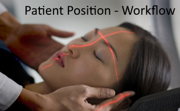
Sup160
2G-RT: Patient Setup & Delivery
This Supplement specifies two IODs to support workflow
management and patient setup for Radiotherapy treatment
delivery sessions. The IODs are designed as part of the
2nd Gen. Radiotherapy object framework.
This Supplement introduces an RT Radiation Set Delivery
Instruction IOD, which specifies the RT Radiations to be
applied for treatment delivery, the order in which they
are applied, and parameters related to the upcoming RT
Treatment Session.
This Supplement introduces an RT Treatment Preparation IOD
to specify setup devices, setup procedures and parameters
related to the setup of the patient prior to the delivery
of therapeutic radiation.
The 1st Gen. RT Plan IOD contained a Patient Setup Module
with a similar content. However, this content may change
between the treatment sessions, while most of the content
of the RT Plan, defining the treatment parameters, remains
the same.
This caused unnecessary proliferation of RT Plan SOP
Instances, and compromised fraction counting within a
series of dosimetrically uniform treatments. Therefore,
the 2nd Gen. RT Radiation Set and RT Radiation IODs use a
separate IOD for this purpose.
Supplement 160 was voted as Final Text and will be
incorporated into the standard with the 2021d publication.
View slideset »
2020
Sup221
Dermoscopy
This Supplement to the DICOM Standard introduces a new IOD
and a new storage SOP Class for encoding and storing dermoscopic
images.
Dermoscopy is a diagnostic technique that enables
visualization of the morphological structures of the
skin. Dermoscopy (also known as dermatoscopy and
epiluminescence microscopy) is a non-invasive, in vivo
skin examination that has demonstrated to be an important
aid in the early recognition of malignant melanoma and
other skin tumors. Dermoscopy is also used for non-skin
cancer disease conditions (e.g., inflammatory disease).
A dermoscope is hand-held device that consists of
magnifier and light source. Emitted light can be polarized light or
non-polarized. Dermoscopic examination can be by direct contact with
skin or noncontact. Dermoscopy using non-polarized light require
direct contact between the skin and the device.
For direct contact dermoscopy an immersion medium is
placed on the skin surface and a glass plate on the dermoscope is
placed directly against the skin. Non-contact dermoscopy does not
require the dermoscope to be in contact with the skin surface.
Three techniques are used in dermoscopy: polarized
non-contact dermoscopy, polarized contact dermoscopy, and
non-polarized contact dermoscopy.
Supplement 221 was voted ready as Final Text. It is
incorporated into the standard in the update "2020e".
View slideset »

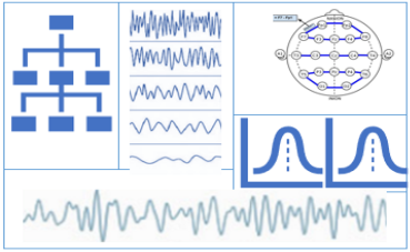
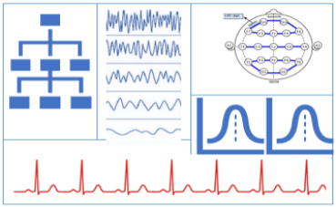
Sup217
Neurophysiology Waveform
This Supplement introduces SOP Classes for storage of neurophysiology
waveforms by adding the related neurophysiology IODs and the necessary
neurophysiology waveform context groups.
This Supplement adds the following SOP Classes:
- To store routine electroencephalography (EEG) data recording the electrical activity of the brain collected on the skull surface using electrode positions of the international 10/10 or 10/20 localization scheme.
- To store electromyography (EMG) data recording the electrical activity of skeletal muscles.
- To store electrooculography (EOG) data collected near the eyes recording eye movement.
- To store electroencephalography (EEG) data acquired during a polysomnography (PSG) study.
- To store respiratory data recorded using more than a single channel.
- To store information about a patient's position continuously.
Supplement 217 was voted into Final Text and to become part of the standard. View details »
View slideset »
Sup208
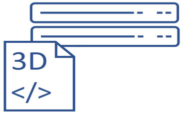
Encapsulated Additional Models 3D Manufacturing
Supplement 208 extends the DICOM Standard to better address
medical 3D manufacturing and uses of Virtual Reality,
Augmented Reality, and Mixed Reality.
These extensions fall in three areas:
- Support for a new 3D model type: Object File (OBJ)
- Identification of models for assembly into a larger object
- Capturing a preferred color for manufacturing or display of a model OBJ Encapsulation
The supplement incorporates not just Object Files (OBJ), and also any supporting Material Library Files (MTL) and texture map files (JPG or PNG) on which an OBJ may rely.
As with Encapsulated STL, the new Encapsulated OBJ, Encapsulated MTL and texture image IODs allow 3D manufacturing models to be exchanged between various types of equipment using DICOM messages. This adds the ability to store, query and retrieve complete OBJ models as DICOM Instances.
Updates are addressed by storing new instances, with reference back to earlier instances in a manner similar to the IOD for STL encapsulation.
This supplement was voted to be ready as final text. It is now being incorporated into the standard. View details »
View slideset »
Sup199
RT Radiation Records
The SOP Classes in this document are defined to record how a
Radio Therapy treatment was performed.
The following IODs are introduces:
- RT Radiation Record Set Storage
- RT Radiation Salvage Record Storage
- Tomotherapeutic Radiation Record Storage
- C-Arm Photon Electron Radiation Record Storage
- Robotic-Arm Radiation Record Storage
This comprises acquired machine values, measured dose values, overrides, etc.
In addition, recording of a manual implementation of a radiation is covered.
This supplement is based on the real-world model and specifications defined in supplement 147. References, definitions etc. not present in this supplement can be found in supplement 147. Additional information is found in Supplement 175 and 176.
Supplement 199 was voted into Final Text and to become part of the standard. View details »
View slideset »
Sup176
2nd Gen. Other non C-Arm RT Treatment
The scope of this supplement is the introduction of new RT
Radiation IODs for non-C-Arm treatment devices.
This supplement adds support for instances of the following RT
devices:
- Tomotherapeutic Radiation
- Robotic Radiation Storage
This supplement was voted to be ready as final text. It is now being incorporated into the standard. View details »
View slideset »
2019

Sup203
Thumbnail Service over DICOMweb
This supplement adds thumbnail handling to web services in
part 18 of the DICOM standard.
This supplement defines Thumbnail resources on the WADO-RS
Study, Series, Instance, and Frame resources in the DICOM
RESTful web services standard. These resources provide
representative images that reflect the content of the parent
resources. The origin server determines the pixel content of
the Thumbnail.
The primary use cases are Thumbnails for image viewers, or
EMR/EHRs, e.g., as referenced from HL7 FHIR Imaging Study
resources.
This resource allows a web client to retrieve a representative
image without having to retrieve a full study structure.
This supplement was voted as Final Text. It will be part of
the next edition of the standard.
Sup202
Realtime Video
This supplement adds realtime video handling to the DICOM standard.
It describes several new DICOM IODs and associated
transfer syntaxes for the transport of real-time video, and/or audio,
and associated medical data. These are referred to collectively as
DICOM Real-Time Video (DICOM-RTV).
The supplement defines an new IP-based DICOM Service for the
broadcasting of real-time video to subscribers with a quality of
service which is compatible with the communication inside the
operating room (OR).
Professional video (e.g., TV studios) equipment providers and users
have defined in SMPTE (ST 2110 family of standards). ST 2110-10 uses a
multicast model rather than a peer-to-peer communication
model. DICOM-RTV builds upon ST 2110.
DICOM-RTV restricts real-time communication to uncompressed video.
This supplement was voted as Final Text. It will be part of
the next edition of the standard.

Sup183
Webservices Redocumentation
This supplement re-documents PS3.18 Web Services.
The goals of this re-documentation are:
- Factor out text that is common to multiple services and in doing so 1) ensure uniformity and 2) make clearer and concise for readers.
- Use a uniform format and style for documenting DICOM web services, making it easier to navigate and more efficient for readers implementing multiple services
- Bring the Standard into conformance with current Web Standards, especially [RFC7230 - 7234], and [RFC3986 - 3987].
- Use the Augmented Backus-Naur Form (ABNF) defined in [RFC5234] and [RFC7405] to specify the syntax of request and response messages.
- Use consistent terminology throughout the Standard.
- Use a consistent format for documenting services and transactions.
This supplement was voted as Final Text and will be part of the next edition of the standard. View details »
Sup175
2nd Gen. C-Arm RT Treatment
This Supplement introduces the RT Radiation Set and representation of the C-Arm techniques.
An RT Radiation Set IOD defines a Radiotherapy Treatment Fraction as a collection of instances of RT Radiation IODs.
Further this supplement adds support for C-Arm Photon-Electron Radiation instances.
RT Radiation IODs represent different treatment modalities.
This supplement was voted as Final Text and will be part of the next edition of the standard.