
Sup252
CT Image Storage for Processing
This Supplement adds For Processing storage SOP Classes based on the
existing CT Image IOD, Enhanced CT Image IOD, and Legacy Converted
Enhanced CT Image IOD.
For Processing storage SOP Classes in DICOM facilitate the exchange
and storage of images needed for processing while distinguishing them
from those for presentation.
This supports appropriate data pipelines while not disrupting reading
workflow with images not intended for presentation.
These new SOP Classes mirror existing Mammography, Intra-Oral X-ray,
and Digital X-ray For Processing SOP Classes.
One application of the For Processing SOP Classes is to store and
exchange CT basis images created by the multi-energy decomposition
process.
These are not typically diagnostic themselves, but can be processed to
generate an extensive variety of diagnostic images (iodine maps,
virtual non-contrast images, virtual monoenergetic images at various
energy levels, calcium maps, etc.).
Hanging Protocols would typically ignore these For Processing
images.
This supplement was voted ready to be incorporated into the standard
as Final Text.
View slideset »
Sup245
RDSR Informative Annex
This Supplement explains the creation and usage of
Radiation Dose Structured Report (traditional and
enhanced) within Angiography, Mammography, Radiography,
Radiofluoroscopy, CT, and Dentistry modalities.
This supplement excludes Radiopharmaceutical Radiation
Dose Structured Report, Patient Radiation Dose Structured
Report, and radiation for treatment (which is encoded in
the family of Radiotherapy objects).
The content definition of the RDSR varies by modality, and
there are many different types of system configurations in
the field.
This supplement provides a clear understanding of the
precise requirements for each type of device.
The purpose of this supplement can be summarized as
follows:
- Give more information beyond the definitions in PS 3.16: describe real-world scenarios of typical equipment configurations, provide examples and encoding guidelines;
- Indicate restrictions on the applicable scenarios (defined terms, value ranges, presence of Content Items);
- Assess the applicability for some conditional Content Items under particular scenarios;
- Promote usage of optional Content Items under particular scenarios;
- Explain similarities and equivalences of same information in both traditional RDSR and enhanced RDSR. Encoding examples of using the traditional RDSR and the enhanced RDSR (introduced in Supplement 214), and mapping between these two RDSRs.
View details »
View slideset »
Sup248
DICOMWeb Send
This supplement adds Send Transactions to DICOMweb’s
Studies and Non-Patient Instances Services to mirror the C-MOVE
operation that is already available in DIMSE.
The Send Transactions have been de- signed with the intention of
facilitating proxies from/to DIMSE.
Furthermore, it defines a C-MOVE option to allow for STOW
sub-operations and an option to allow for C-MOVE SCP proxies,
delegating to a DICOM- web Send origin server.
Security plays an important role for this supplement, as this
supplement defines the three party DICOM- web Send Transaction, and
provides means to cross two DICOM protocols, i.e., between DICOMweb
and DIMSE, at both the operation level and the sub-operation level.
Yet, DICOM considers security orthogo- nal to functionality, and hence
applicable security mechanisms and considerations are described in a
sep- arate supplement.
This supplement will be further presented to and discussed
with WG-06 before going out for Letter Ballot.
View slideset »
Sup250
2D Total Body Photography
This supplement introduces Total Body Photography (TBP) Regional Image
Information Object Definition (IOD) for imaging of a skin
region.
TBP aims to image a person’s entire skin surface. TBP is predominantly
used for sequential screening for skin cancer, in particular melanoma,
but can also be used for the assessment of inflammatory skin diseases
such as psoriasis.
TBP may be 2D or 3D. This supplement addresses 2D TBP. 2D TBP produces
2D planar images, whereas 3D TBP produces volumetric images.
TBP uses visible light imaging. Camera movement may be manual or
automatic. There are typically multiple image acquisitions with the
patient in different positions. Multiple regional images are acquired
to image entire skin surface. TBP is often used in combination with
dermoscopy.
This supplement was voted ready to go out for Public Comment review and voting.
View slideset »
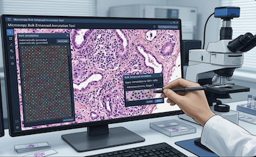
Sup255
Microscopy Bulk Enhanced Annotation
This supplement will add enhancements to the annotation
mechanism in microscopy beyond the support already
available.
This supplement adds the Microscopy Bulk Enhanced
Annotation SOP Class to extend the capabilities of the
existing bulk Microscopy Bulk Simple Annotation IOD to
enable the definition of annotations that define
overlapping vector graphics with mutually exclusive
meaning.
Examples of annotations that this proposal will enable
include: heatmaps, holes/voids, and other
nested mutually exclusive concepts, e.g., tumor tissue
contained within healthy tissue.
The Microscopy Bulk Simple Annotation SOP Class and IOD
(Supplement 222) were added to the DICOM Standard to
provide a mechanism to efficiently encode machine learning
(ML) and human generated vector graphics annotations for
slide microscopy imaging. The Microscopy Bulk Simple
Annotation IOD supports vector graphic definitions of
points, lines, polylines, rectangles, ellipses, and
polygons.
This supplement will be further presented and discussed
before going out for Public Comment.
View slideset »
Sup254
Digital Signature and Media Storage Security
This Supplement updates DICOM Digital Signatures and Secure Media
Storage by adding support for modern algorithms while maintaining
backward compatibility.
In recent years, regulations concerning security and privacy have
become increasingly prominent across countries and regions (e.g., EU
GDPR, US FDA Cybersecurity Guidance, China Cybersecurity Law, Data
Security Law and Personal Information Protection Law).
These require strengthened confidentiality, integrity, and
authenticity of medical data.
The Digital Signature Profiles (PS3.15 Section 6.3 and Annex C) and
Media Storage Security Profiles (PS3.15 Section 6.4 and Annex D)
contain legacy cryptography technologies that are still being used,
e.g. 3DES and SHA-1.
This supplement adds support of the newer cryptography algorithm
e.g. ECDSA/EdDSA for digital signature, cryptography technologies
such as authenticated encryption (e.g. AES-CCM/GCM), and key
management technologies such as KEM.
This supplement includes:
- Add 4 new RSA-based Digital Signature Profiles by deprecating legacy algorithms such as SHA-1 and add newer algorithms such as SHA-3 family and upgrade certificate to X.509 V3.
- Add 4 new Digital Signature Profiles which are based on Elliptic Curve algorithms.
- Allow implementations to claim conformance as signer/verifier separately. Implementations conforming to these newer profiles might generate signatures with new algorithms as signer, but meanwhile support deprecated algorithms as verifier to keep backward compatibility.
- Add one new Media Storage Security Profile, which supersedes the current mechanism in Basic Media Storage Security Profile, but use new algorithms, and add support of more key management technologies including Key Agreement (e.g., ECDH) and Key Encapsulation Mechanisms (KEM).
- Add one new Media Storage Security Profile using CMS Authenticated-Enveloped-Data with Authenticated Encryption (e.g., AES-GCM, AES-CCM).
View details »
View slideset »
Sup253
Waveform Compression
This supplement proposes to:
- Add an encapsulation mechanism for DICOM waveforms to encapsulate compressed data in analogy to the existing image compression and encapsulation of compressed pixel data.
- A new Transfer Syntax for lossless compressed DICOM waveforms is added.
- A new Transfer Syntax for lossy compressed DICOM waveforms is added.
This supplement adds Transfer Syntaxes for compression of DICOM Waveforms based on ITU/VCEG codec T.261, which was created especially for encoding and decoding of biomedical waveform data.
This implies the introduction of an encapsulation mechanism for waveform sample data.
T.261 specifies an interoperable format for efficient compression, transmission, and storage of waveform signals, including biomedical waveform signals such as electrocardiography (ECG), electroencephalography (EEG), electromyography (EMG), and photoplethysmogram (PPG) data, as well as other types of general waveform data. It supports various bit depths, a broad range of sampling rates, and large numbers of channels, as required by a range of biomedical and general signal processing applications. T.261 provides for lossy, near-lossless, and lossless compression, and includes features such as optimized data blocking, indexing for rapid access and independent channel decoding, and metadata support. A draft version of the specification of T.261 is available here: VCEG-BZ02-H Spec.
This supplement will be further presented and discussed before going out for Public Comment.
View details »
View slideset »
Sup256
Live Streaming in DICOMWeb
This supplement adds functionality to DICOMweb which enables live
streaming to DICOMweb in a standardized way.
This supplement was voted ready to get a supplement number and will be
further presented and discussed in the base standard group before
going out for Public Comments.
View slideset »